Corneal diseases encompass a range of conditions affecting the transparent front part of the eye, called the cornea. The cornea plays a crucial role in focusing light onto the retina, enabling clear vision. Numerous factors can contribute to corneal diseases, but the primary cause often varies depending on the specific condition.
One of the main causes of corneal diseases is infections, which can be bacterial, viral, fungal, or parasitic in nature. These infections may result from poor hygiene, contact lens misuse, or exposure to contaminated water or foreign bodies. Bacterial keratitis, viral keratitis (such as herpes simplex virus or varicella-zoster virus), and fungal keratitis are common examples.
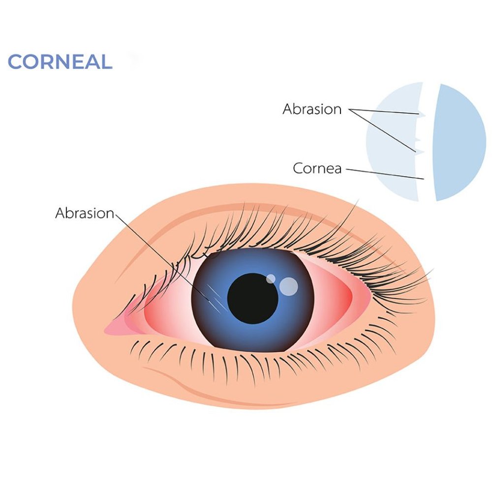
Another significant cause of corneal diseases is trauma, which can range from minor scratches to severe injuries. Trauma may occur due to accidents, foreign objects entering the eye, or surgical complications. These incidents can lead to corneal abrasions, lacerations, or even more serious conditions like corneal ulceration or perforation.
Furthermore, certain systemic diseases such as diabetes, autoimmune disorders, and genetic conditions can predispose individuals to corneal abnormalities. These conditions may affect the cornea directly or indirectly through associated complications like dry eye syndrome or inflammation.
Environmental factors like prolonged exposure to ultraviolet (UV) radiation, pollutants, and allergens can also contribute to corneal diseases. Chronic exposure to these elements can lead to conditions such as pterygium, photokeratitis, or allergic keratoconjunctivitis.
Additionally, degenerative disorders like keratoconus, where the cornea gradually thins and bulges outward, are significant causes of corneal impairment. Genetic predisposition and environmental factors may play roles in the development of such conditions.
You can protect your eye health and potentially slow the process of cataracts by: Not smoking, Protecting your eyes from the sun, Getting regular eye care, and Wearing sunglasses and a hat with a brim to block ultraviolet sunlight.
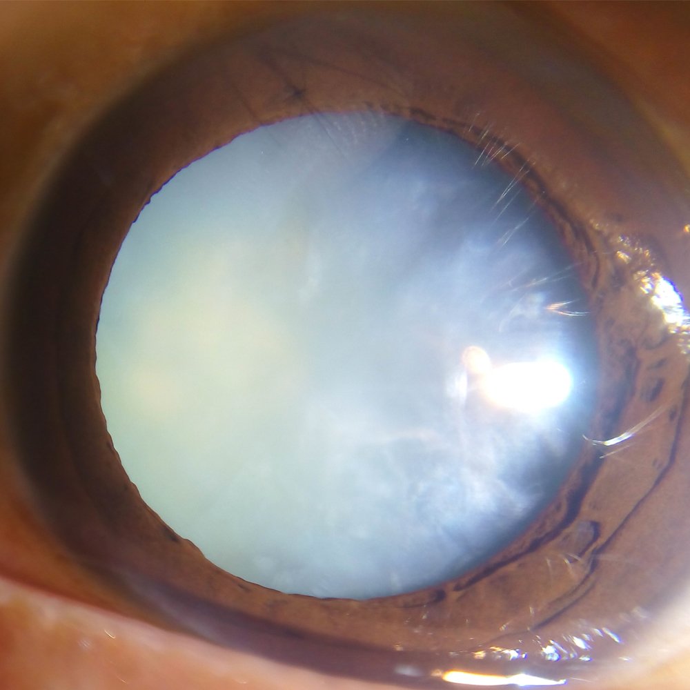
Corneal disease, also known as ocular surface disease, is a group of serious conditions that can affect the cornea. These conditions can cause the cornea to become distorted, clouded, or scarred, and can even lead to blindness.
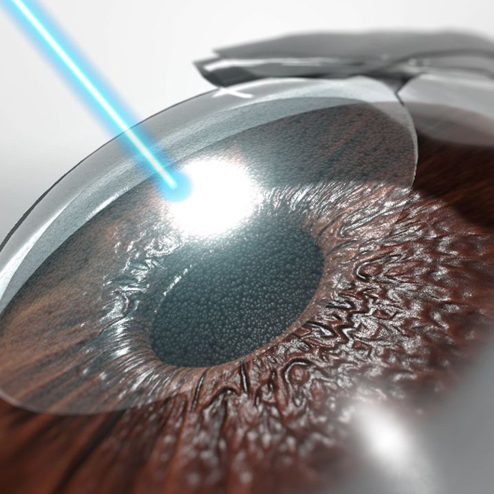
Refractive surgery is an optional eye procedure that improves the eye's refractive state and can reduce or eliminate the need for glasses or contact lenses . It can involve reshaping the cornea, implanting a lens, or replacing the lens.

The retina is the light-sensitive layer at the back of the eye that receives light and converts it into chemical energy. The uvea is the middle layer of the eye between the retina and the sclera (white part of the eye). The uvea is made up of three parts !

Emphasizes fruits, vegetables, whole grains, and fat-free or low-fat milk and milk products.

There are many variations of passages of Lorem Ipsum available, the majority have suffered alteration

There are many variations of passages of Lorem Ipsum available, the majority have suffered alteration
Refractive surgery can correct refractive errors like nearsightedness, farsightedness, astigmatism, or presbyopia. Some of these surgeries reshape the cornea. Others implant a lens in your eye. Either way, the goal is the same. These surgeries focus light correctly on the retina so you can see more clearly.
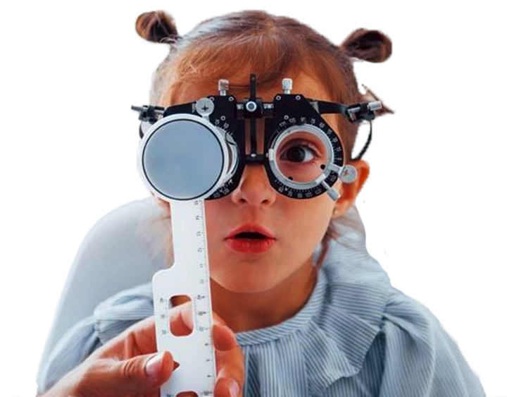
Pediatric ophthalmology is a subspecialty of ophthalmology that concentrates on treating the various eye problems affecting children. Studies show that a lot of Attention Deficit Hyperactivity Disorder (ADHD) and learning issues in children can be attributed to vision problems.


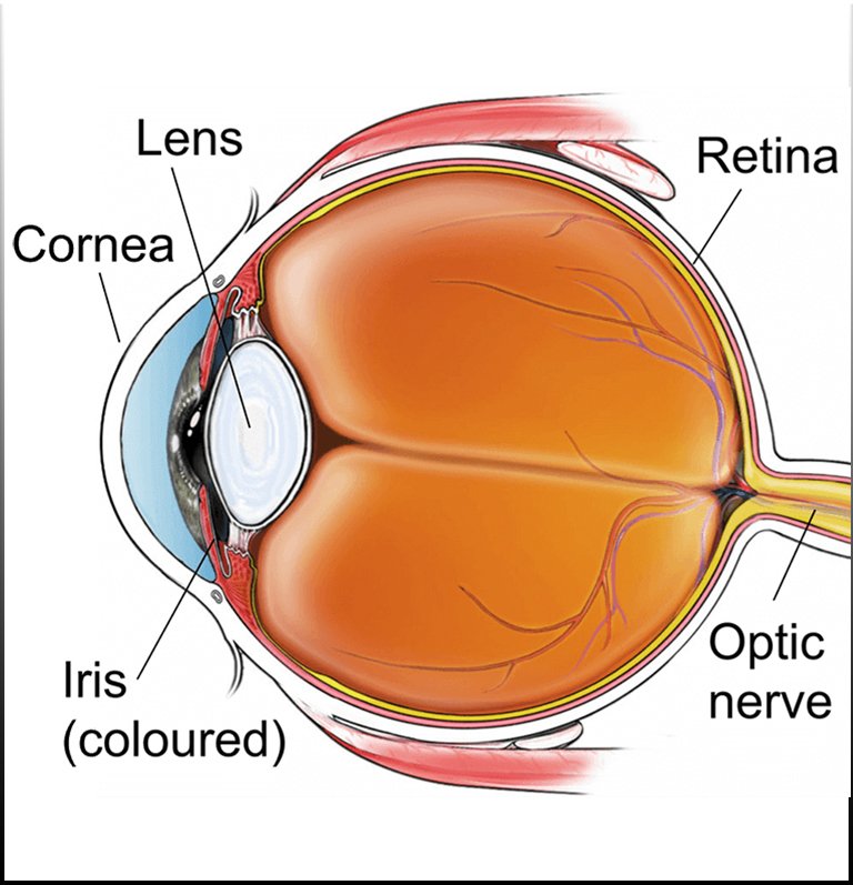
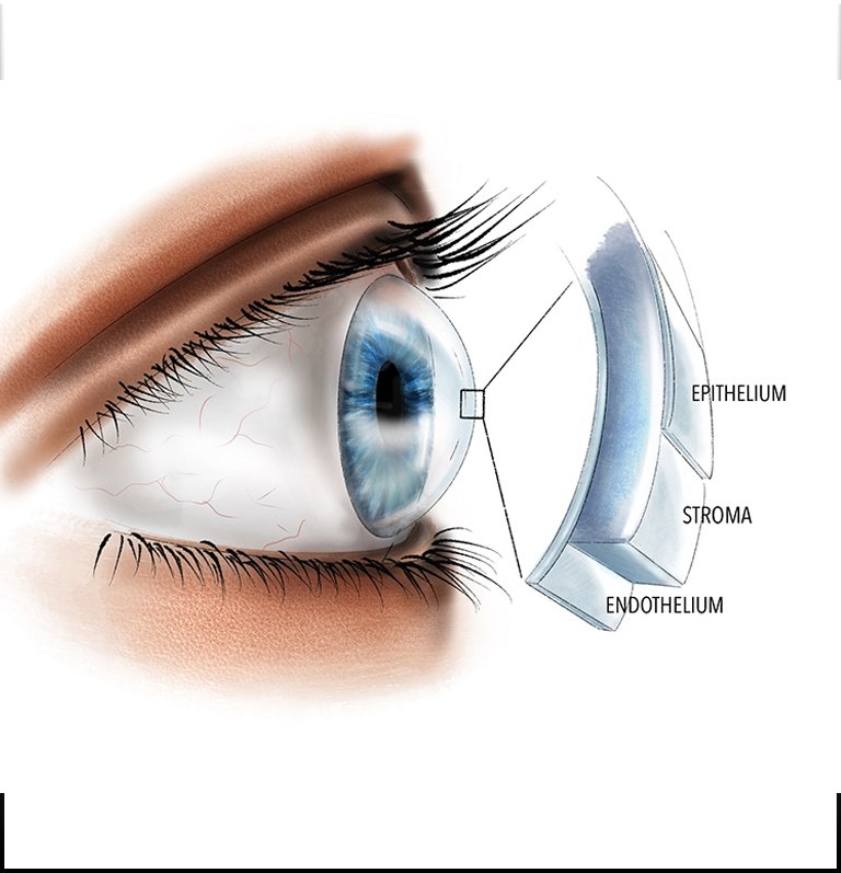
The cornea is the transparent, dome-shaped layer covering the front of the eye. It plays a crucial role in focusing light onto the retina, enabling clear vision. The cornea starts its formation during embryonic development, around the fourth week of gestation. It originates from the outer layer of the embryonic tissue called the ectoderm.
As the embryo develops, specialized cells within the ectoderm differentiate into the cells that will eventually form the cornea. These cells undergo a process called epithelial-mesenchymal transition (EMT), where they transform from epithelial cells, which form the surface of the body, into mesenchymal cells, which are multipotent and can differentiate into various cell types.
During this transformation, the mesenchymal cells migrate and aggregate in a specific area in front of the developing eye called the ocular surface ectoderm. Here, they begin to proliferate and differentiate into different cell types that will contribute to the structure of the cornea, including stromal cells, endothelial cells, and keratocytes.
Simultaneously, a thin layer of epithelial cells covers the surface of the developing cornea, forming the corneal epithelium. This epithelial layer serves as a protective barrier and helps maintain the transparency of the cornea.
Throughout fetal development and into infancy, the cornea continues to grow and mature, achieving its final shape and optical properties. After birth, the cornea undergoes minimal changes in size and shape but remains a dynamic structure capable of regeneration and repair throughout life.
Overall, the formation of the cornea is a complex process involving the orchestrated differentiation and migration of cells derived from the embryonic ectoderm. This intricate development ensures the proper structure and function of the cornea, essential for clear vision.